Kidneys that might be considered...suboptimal...for a number of reasons are sometimes subjected to something called NMP that is in the blog title and I doubt i can type it correctly again.
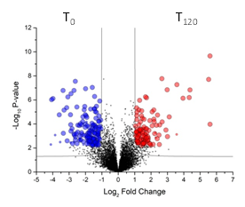
As you might guess, having human kidneys in a machine to keep them going for a transplant imparts a lot of changes. Knowing what ones might help us predict which will work, which won't, and how to preserve them longer!
I can't figure out what instrument was used for this. Since the MS1 resolution is 120,000, it is probably a high field Orbitrap, and I suspect we're operating in DDA mode. The biology is the star here and the details on how the proteomics was performed doesn't fit within the page limits. That's still okay, because of how interesting the biology is.
If you actually want to know, the RAW files are up on ProteomeXchange here as PXD036432.


ReplyDeletefor those interested in experimental conditions:
Analysis of renal tissue peptides by nano-UHPLC-MS/MS
Peptides were separated on a 200-cm µPAC C18 column (PharmaFluidics) mounted in a thermostatic column compartment maintained at 50 °C. We applied a gradient of 5–7% buffer B (80% acetonitrile in water, supplemented with 5% DMSO and 0.1% formic acid) at a flow rate of 750 nl/min dropping to 350 nl/min over a period of 12 min. The peptides were then eluted in a non-linear gradient of 7–45% buffer B at a constant flow rate of 350 nl/min for 78 min. The fractions were analyzed in data-independent acquisition (DIA) mode, with Orbitrap detection for MS1 at a resolution of 120,000 within the range 375–1500 m/z and an AGC target of 300%. Advanced peak determination was enabled for MS1 measurements. The FAIMS compensation voltage was set to –50 V at standard resolution. Precursors were selected for data-independent fragmentation in 60 windows of 380–980 m/z with an overlap of 2 m/z. The higher-energy C-trap dissociation (HCD) value was set to 30% and MS2 scans were acquired at a resolution of 15,000 with a maximum injection time (max. IT) of 22 ms and an AGC target of 1000%.
Raw data were processed using FragPipe v17.1 with MS Fragger v3.452 in the DIA_SpecLib_Quant workflow53 to build spectral libraries, and DIA-NN v1.8 for DIA quantification. All default settings were applied except the RT Lowes fraction, which was modified to 0.01 for spectral library generation. We screened the UniProt human protein sequence database (release UP000005640_9606 July 2021) and a database of common contaminant proteins.
Analysis of urine peptides by nano-UHPLC-MS/MS
Peptides were separated as above, but using a 1–7% initial gradient of buffer B. The fractions were analyzed in data-dependent acquisition (DDA) mode with the same resolution and m/z range as stated above for the analysis of renal tissue proteins. The top 10 precursors were selected for MS2 analysis. MS/MS spectra were acquired in the linear ion trap (rapid scan mode) after HCD with a collision energy of 30% and a custom AGC target. We applied quadrupole isolation with a 1.6 m/z window, dynamic exclusion set to 50 s, and a max. IT of 35 ms.
Raw data were processed using MaxQuant v2.0.3.054. A false discovery rate of 0.01 was used for the identification of proteins, peptides and peptide-spectrum matches. A minimum of seven amino acids was required for peptide identification. The Andromeda engine in MaxQuant was used to search MS/MS spectra against the UniProt human database (release UP000005640_9606 July 2021). N-terminal acetylation, methionine oxidation and asparagine/glutamine deamidation were selected as variable modifications, and carbamidomethyl cysteine was selected as a fixed modification. Quantification intensities were calculated using the default fast MaxLFQ algorithm with the activated option “match between runs.” Raw data processed by MaxQuant were further processed using IceR55 with default settings, to reduce the number of missing values.